

2019 General Electric (GE) Signa Explorer 1.5T

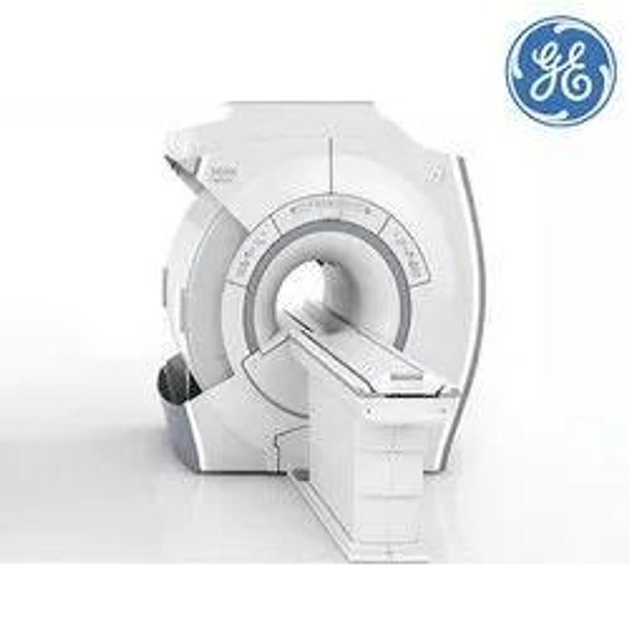
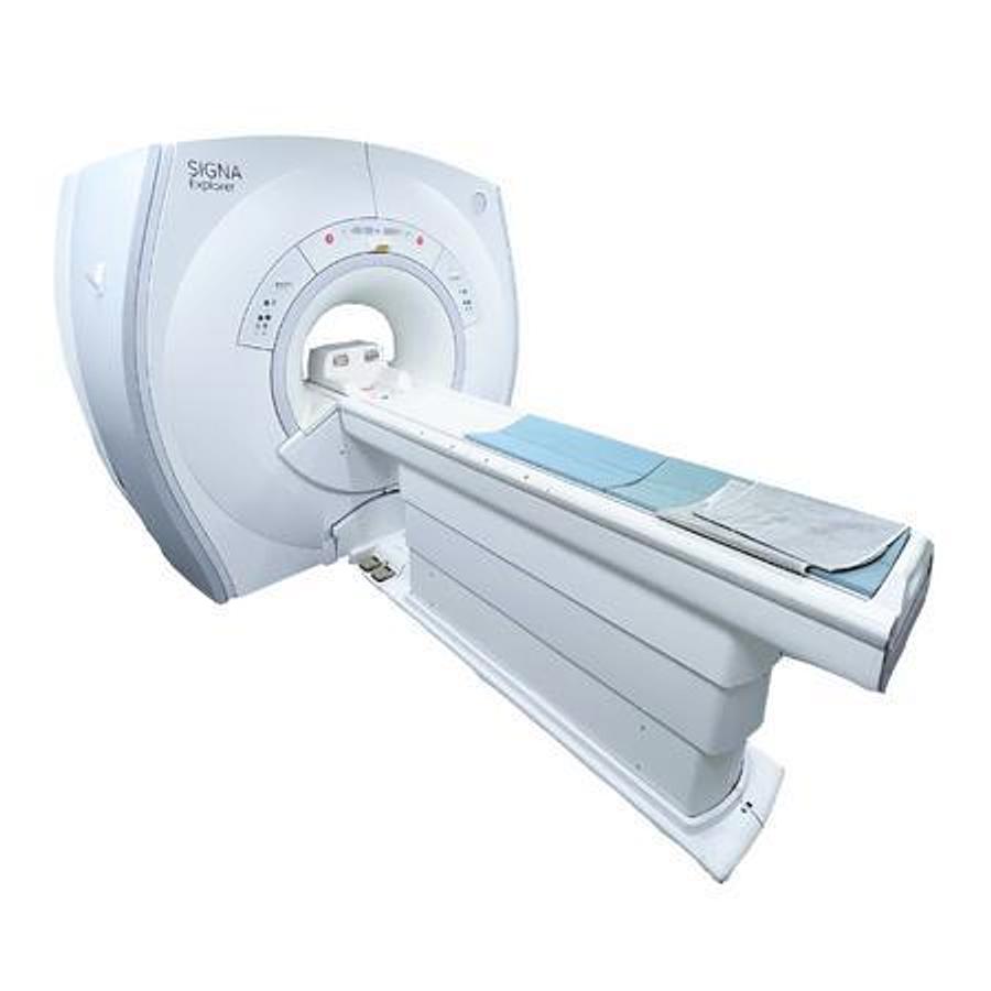

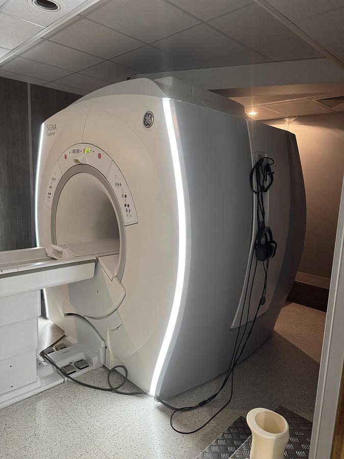
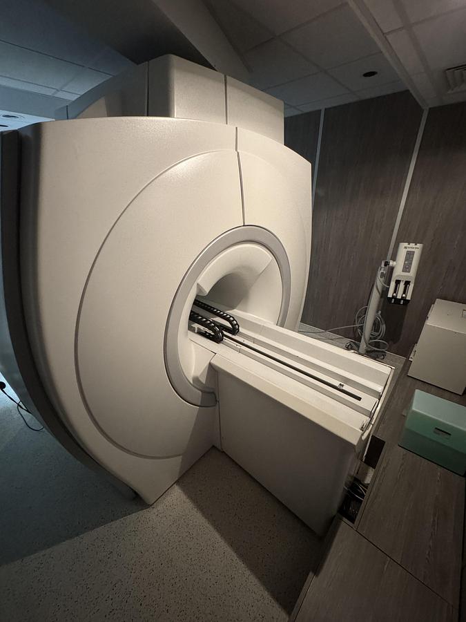
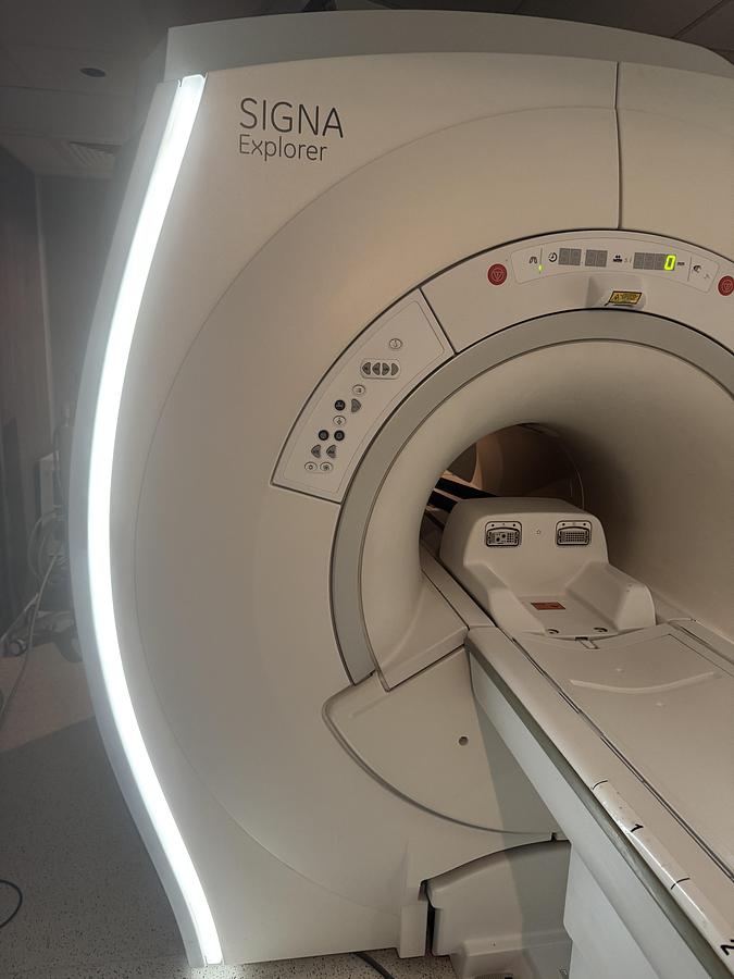

2019 General Electric (GE) Signa Explorer 1.5T
Description
Magnet Features and Gradients:
The 1.5T SIGNA Explorer system features a modern, wide-bore LCCw superconducting magnet that is stable and highly homogeneous. The magnet weighs 3900 kg and is 195 cm long.
- Homogeneity of the magnet:The high homogeneity of the magnet contributes to high image quality in applications such as large field of view (FOV) imaging, up to 50cm×50cm×50cm, and robust and reliable fat saturation.
- Gradients:The gradients offer exceptional spatial and temporal resolution. They have a peak amplitude of 33 mT/m and a peak slew rate of 120 T/m/s5. They are designed to be non-resonant and actively shielded to reduce eddy currents.
- Magnet cooling:The magnet is cooled solely by liquid helium and features a "zero boil" function under normal operating conditions.
- Acoustic noise reduction:The system includes a special vibroacoustic damping pad to isolate the magnet from the building, reducing the transmission of acoustic noise to surrounding structures. It also features ART (Acoustic Reduction Technology) to modify pulse sequences and reduce acoustic noise without compromising image quality.
Patient Table and Comfort:
The system can be configured with one of two patient table options:
- Low height fixed table:It rises from 49.0 cm to 96.5 cm and supports a maximum weight of 200 kg (440 lbs) for scanning.
- Removable table:Allows the technologist to prepare a patient outside the scanning room while another is being scanned. In the event of an emergency, it allows the patient to be removed from the scanning room in less than 30 seconds. The standard detachable table and the "Lite" have a maximum scanning weight of 160 kg.
- Patient comfort features:The magnet design includes a dual flared hole, lighting and ventilation inside the hole, a two-way intercom, and feet-first positioning.
Processing Hardware and Software:
The system is equipped to handle demanding applications thanks to its processing speed and storage capacity.
- Host computer:It uses an Intel Xeon W-2123 CPU clocked at 3.6 GHz and 64 GB of main memory. It can store 3,300,000 uncompressed 256×256 images.
- Rebuild Engine:It features a Dual Intel Xeon Silver 4110 processor with 64GB of memory and can reconstruct 37,000 FFTs/second.
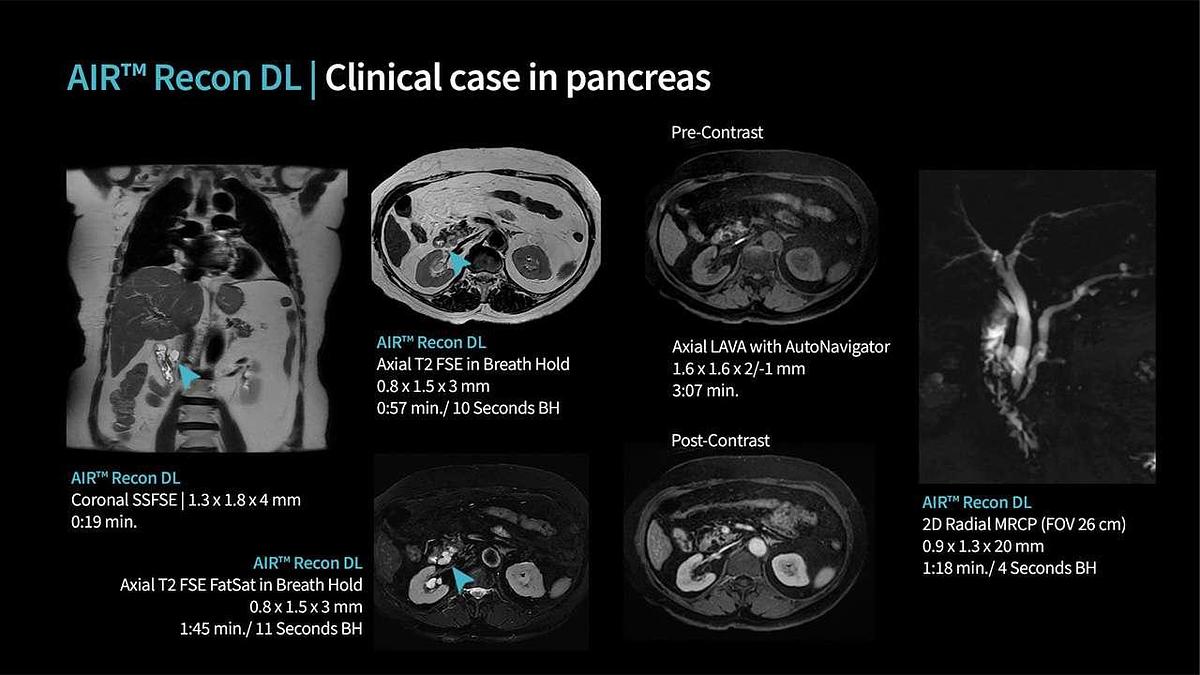
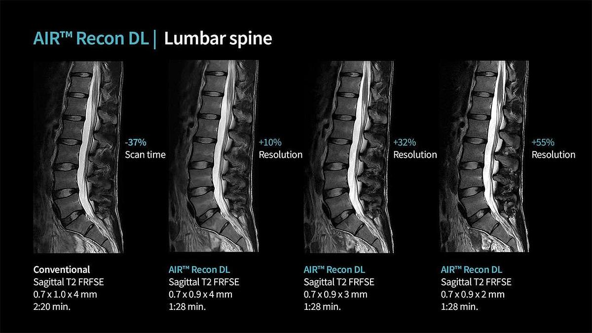
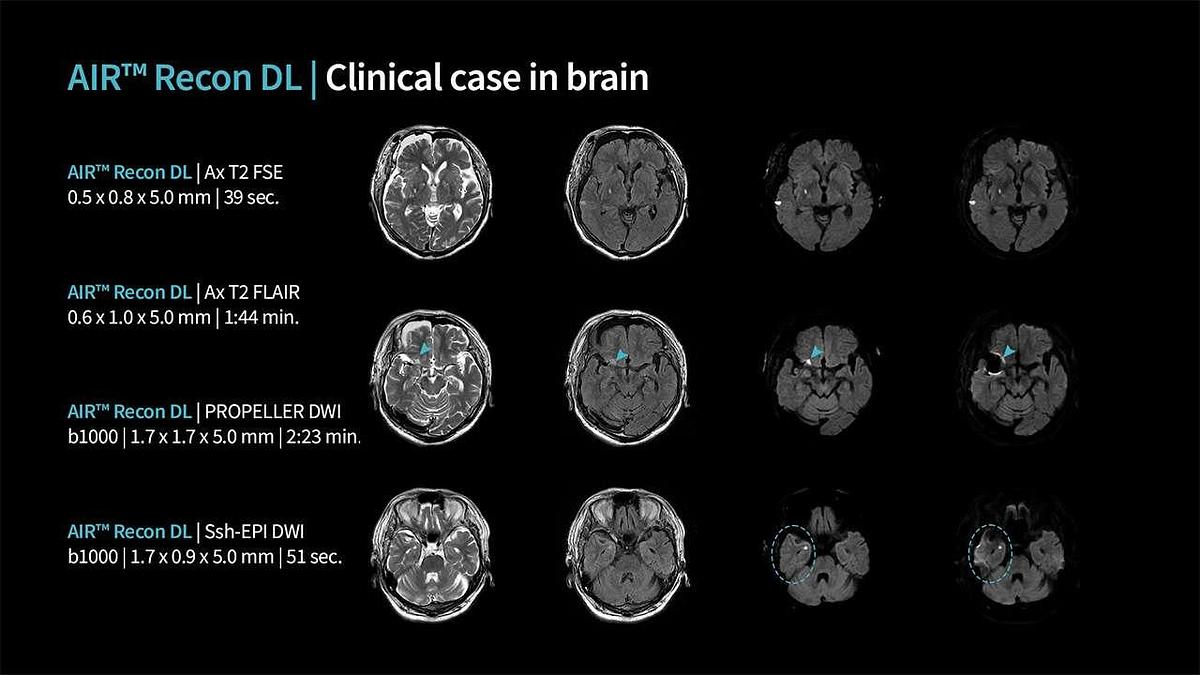
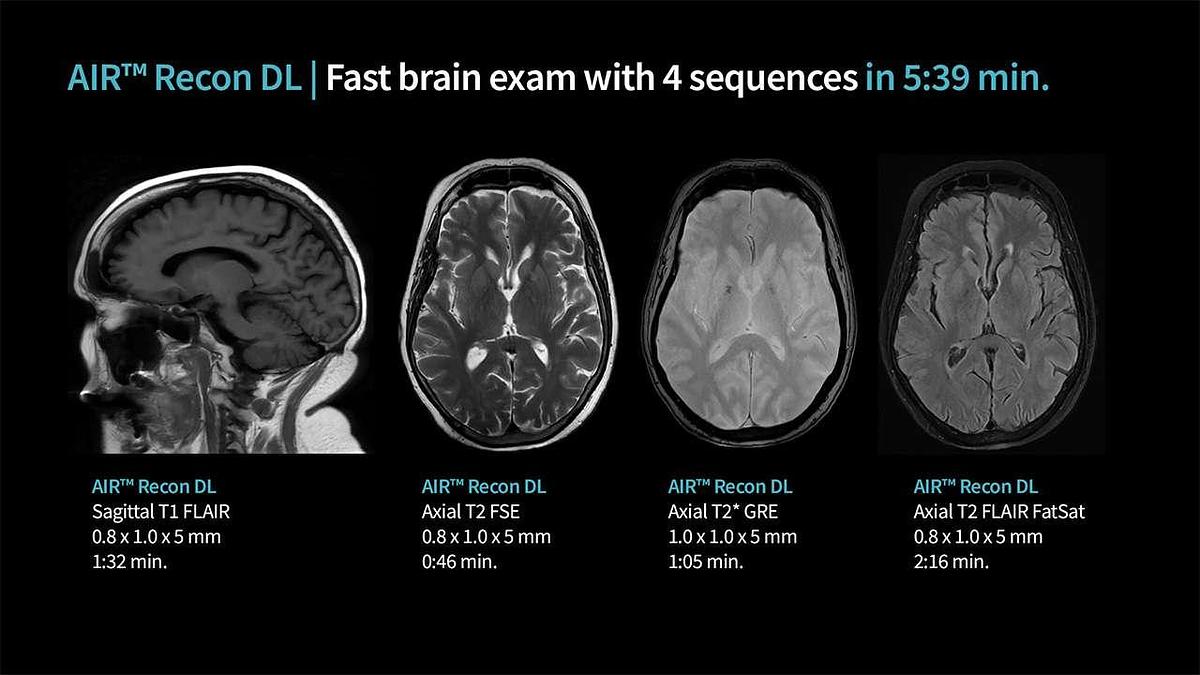
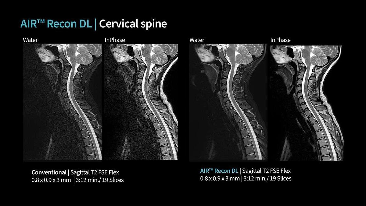
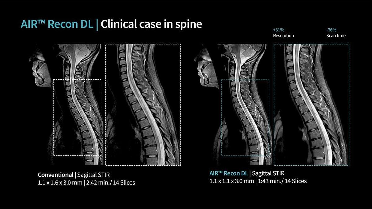
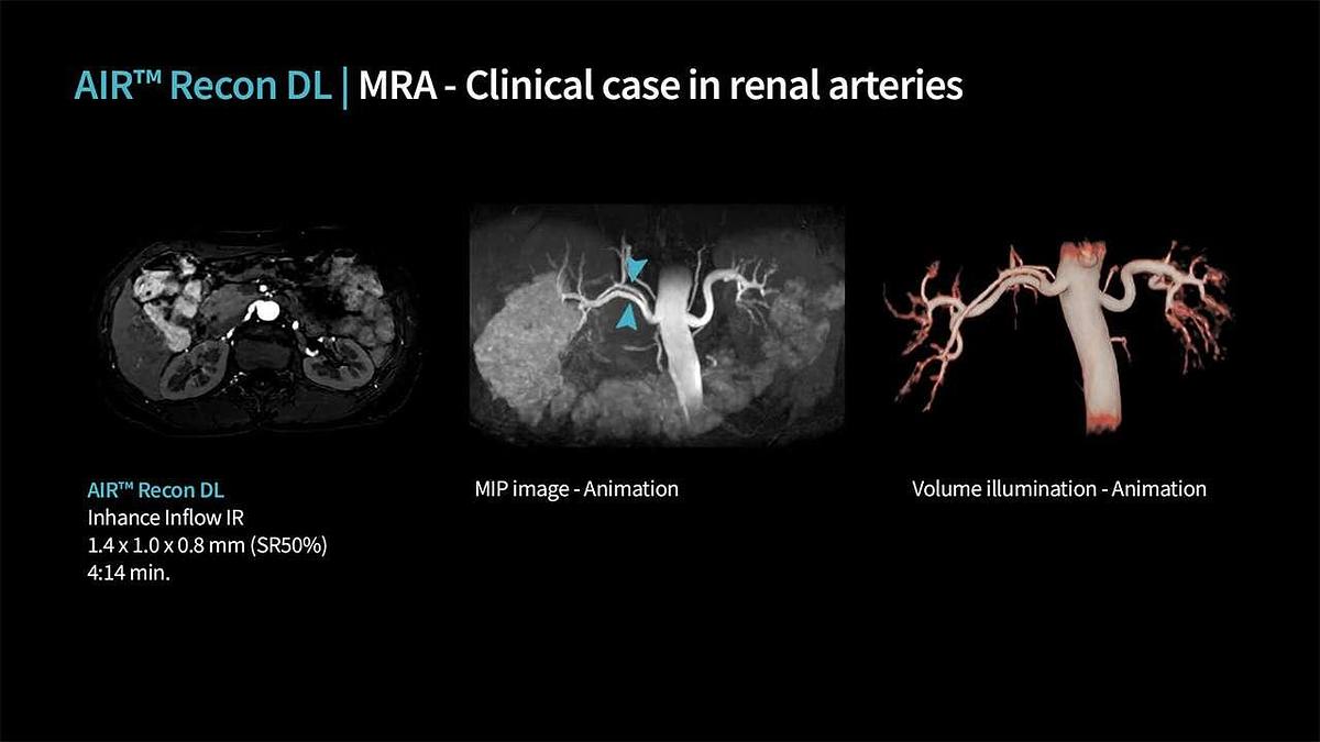
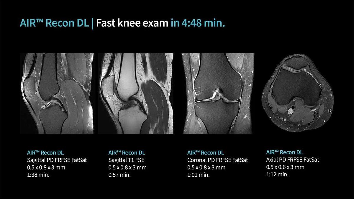
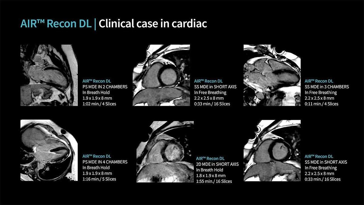
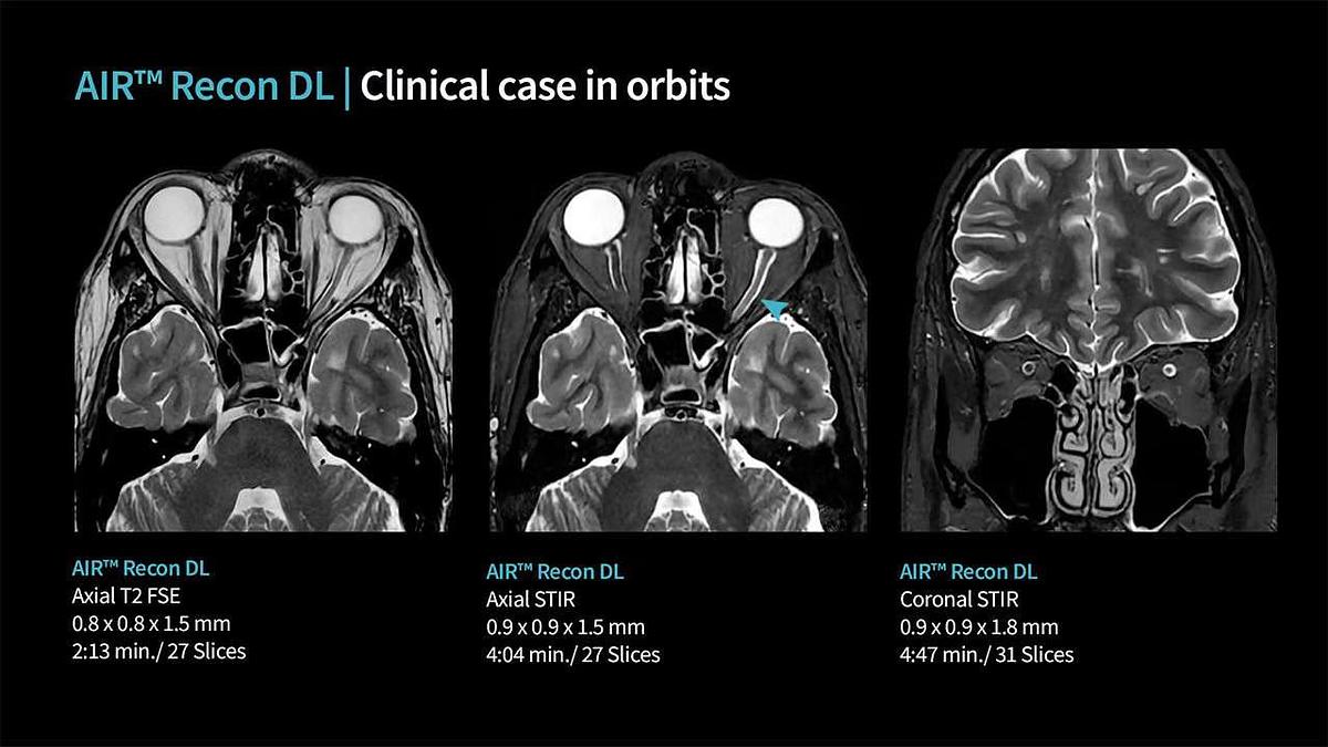
- Deep learning reconstruction:The AIR™ Recon DL application uses trained neural networks to remove noise and artifacts from the reconstructed image, improving the signal-to-noise ratio (SNR) and sharpness, which can enable shorter scan times.
- Updated console: Permanent cardiology options enabled.
- DICOM 3.0
Workflow:
SIGNA Explorer’s AutoFlow suite is designed to make your workflow easier and more efficient.
- Ready Brain:Automates brain examination steps, from acquiring a localization image to transferring the final data, resulting in greater consistency.
- Auto Protocol Optimization (APX):Enables a simple, automated workflow for breath-hold imaging.
- Inline Processing:It automates many routine tasks that previously required user interaction, automatically completing processing steps after data reconstruction.
- Linking:Automates image prescription for each series in an exam, combining information from one prescribed image series with all subsequent series.
- AutoVoice:It offers pre-recorded, user-selectable instructions in more than 14 languages, helping to guide the patient consistently throughout the exam, especially in studies requiring breath-hold monitoring.
Image and Reel Options:
The system is compatible with a variety of imaging techniques and coils.
- Parallel images:Includes techniques such as ASSET and ARC to accelerate data acquisition.
- Dynamic images:Supports LAVA and LAVA Flex for high spatial and temporal resolution torso imaging. It also features QuickSTEP for automatic multi-station image acquisition and blending for peripheral vascular studies.
- Advanced techniques:Includes MR Touch for measuring relative tissue stiffness, PROSE for prostate lesion assessment, SWAN 2.0 for visualizing iron deposits and small vessels, and IDEAL IQ for water and fat separation.
- RF coils:The system includes an integrated head coil and body coil. Optional coils are also available, such as Flex Arrays and dedicated coils for breast, shoulder, heart, knee, foot/ankle, and wrist.
Permanent options enabled:
ARC, 3D ASL, Asset, Blood Flow and Volume Measurement, Bloodsupp,
BRAVO, BREAST2, Cinema, CINE IR, COSMIC, Cube T2, 3D Dual Echo, 3D
Delayed Enhancements, DW EPI, E3DTOF, Enhanced DWI, Echo Planer
Imaging, Fastcine, Fast Gradient Echo, Fiesta 2D, 2D Fat SatFiesta, Fiesta 3D,
3D Fat Sat FIESTA, FIESTA-c, FLAIR3D, FLAIR EPI, Flow Analysis, 3DFRFSE,
Fast Spin Echo and FLAIR,FSE_XL, Fluoro-triggered MRA, Time of Flight, 3D
Heart, Modality Worklist, IDEAL, iDrive, iDrive Pro, Inhance Deltaflow,
Inhance 2D Inflow, Inhance 3D Velocity, Inhance 3D Inflow IR, Lava, LAVADE,
LAVA-XL, 2D MERGE, 3D Merge, multi-echo fgre, Multi-Phase(variable
delays), Navigator, Phase Contrast Vasculat Imaging, Performed Procedure
Step, ProbePRESS, PROPELLER, DW PROPELLER, T1 Flair PROPELLER, T2
PROPELLER, T2 Flair PROPELLER, QuickSTEP, iDrive Pro Plus, Smart Prep,
SPECIAL, SSFSE, SSFSE MRCP, T2Star Weighted Angiography, T2MAP,
Diffusion Tensor, Three Plane Localizer, FiberTrak, TRICKS, VIBRANT-DE, IP
Protection, IDEAL IQ, eXtremePerformance Gradient,Phase Imaging
Technique, MAVRIC SL, Express Spine Annotation, Body Navigator,
Prospective Motion Correction, Silent MR, Focus, Silent Propeller, DW Prep,
DISCO, Chemical Shift, Cube DIR, SIGNA Explorer, Auto Navigator Tracker,
Auto Protocol Optimization, Synthetic DWI, Flex, Hypersense, HyperCube,
Intergated Registration, Volume Viewer API, Cube STIR, WB DWI, Brain
View, Body View, CARDIAC 3D.
Specifications
| Manufacturer | General Electric (GE) |
| Model | Signa Explorer 1.5T |
| Year | 2019 |
| Condition | Refurbished |
| Stock Number | 0067 |
| Magnetic field (T): | 1.5T |
| Gantry ring diameter: | 60 cm |
| Number of channels: | 16 channels |
| Coils included: | Shoulder, knee, head, body, foot, ankle, wrist, large flexion and small flexion. |